Understanding Minimally Invasive Lumbar Diskectomy
Lumbar diskectomy is a type of surgery to remove a part of a disk in the lower back. Minimally invasive surgery uses two or more small cuts (incisions) instead of one large cut. This may lead to less pain after surgery. It can also lead to faster recovery.
Parts of the spine
Your backbone (spinal column) is made up of a chain of bones called the vertebrae. Your spinal cord runs through the spinal column. The bones help protect the cord from injury. Disks sit between each vertebra. The disks give cushioning and support. Large nerves called nerve roots lead from the spinal cord through small holes between the bones. These holes are called foramen. These nerve roots send and receive signals to and from the body. The signals are sent to and from your brain through the spinal cord.
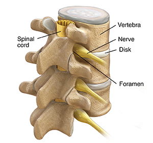
The outer wall of one of these disks may dry out and weaken with age or injury. When this happens, the soft inner part of the disk bulges out. This is called a herniated or bulging disk. This bulging disk can press on the spinal cord or the nerve roots. This can cause pain, tingling, difficulty urinating, or weakness in your arms or legs.
Why minimally invasive lumbar diskectomy is done
You may need this surgery if you have a herniated disk in your lower back that is causing symptoms. Symptoms may include weakness, pain, or tingling in the back area and in one of your legs.
Lumbar diskectomy can’t be used to treat all cases of back pain. And not everyone with a herniated disk needs this surgery. Your doctor might advise the surgery if you have tried other treatments but still have severe symptoms. Other treatments to try first include physical therapy and anti-inflammatory medicines.
How minimally invasive lumbar diskectomy is done
The surgery is done by a neurosurgeon or an orthopedic surgeon and a trained medical team. The surgeon will use a special type of X-ray to view the surgery. The doctor will make a small incision on your back in the area that needs to be treated. The surgeon will put a tool called a tubular retractor into this incision. This will expose the part of the spine to be treated. The surgeon will then pass small tools through this retractor. This may include a tiny camera and a light. Your doctor will remove the herniated part of the disk using small tools. Other repairs will be done as needed.
Risks of minimally invasive lumbar diskectomy
Every surgery has risks. Risks for this surgery include:
Not everyone improves with surgery. Your risks may vary depending on your age and your general health. Talk with your doctor about the risks that most apply to you.
Online Medical Reviewer:
Anne Fetterman RN BSN
Online Medical Reviewer:
Luc Jasmin MD
Online Medical Reviewer:
Raymond Kent Turley BSN MSN RN
Date Last Reviewed:
12/1/2022
© 2000-2024 The StayWell Company, LLC. All rights reserved. This information is not intended as a substitute for professional medical care. Always follow your healthcare professional's instructions.