Having Carotid Artery Stenting
Understanding carotid artery stenting
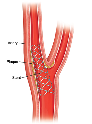
Carotid artery stenting is a procedure to open up a narrowed carotid artery. Two carotid arteries on each side of the neck deliver blood to the brain. A stent is a tiny, metal mesh coil that props open the artery so blood can flow freely. During the procedure, your doctor puts a long thin tube (catheter) into an artery. This lets the doctor move tools through the artery to put the stent in place. Stenting is often done with a procedure called angioplasty. For angioplasty, the doctor inflates a tiny balloon at the tip of the catheter at the blocked part of the artery. The inflated balloon presses the plaque against the artery wall. This opens the artery for better blood flow. The doctor then places the stent to help keep the artery open.
Why is carotid artery stenting done?
Carotid artery stenting is done to restore blood flow to the brain to prevent stroke. A stroke can occur if the artery becomes narrowed. Or if a blood clot forms in the artery. This can block blood flow to part of the brain.
How is carotid artery stenting done?
Your doctor will need to talk to you during the procedure, so you’ll be awake the entire time.
Putting in the catheter
-
A surgical team member will clean the skin in the area of the insertion site with an antiseptic liquid. Your doctor will numb it with local anesthesia.
-
The doctor will make a puncture. It's usually in the femoral artery in the groin. Or they may use an arm or even neck artery.
-
The doctor will put an introducer sheath or tube into the puncture.
-
The doctor puts the catheter into the sheath. Using X-rays as a guide, the doctor moves the catheter through the sheath and up through the aorta and into the carotid artery.
-
You will have an angiogram of the carotid artery. A carotid angiogram is a test that uses X-rays and a special fluid (contrast medium or dye). It helps your doctor find blockages or other problems in the carotid arteries. Your doctor injects the contrast dye into the artery to show where the narrowed portion is on X-ray. You may feel warmth toward your head just after the dye is injected. You may also think you see some flashing lights. This is normal and will only last for a few seconds. Let your doctor know right away if it continues.
Placing the filter
A filter or other protective device prevents pieces of plaque from being carried downstream into the brain and causing a stroke. The doctor uses the catheter to place the unopened filter in the artery. They move the filter past the narrowed area. They then open the filter. It stays in place for the whole procedure. If narrowing is very severe, your doctor may need to widen the artery before the filter is put in place.
Opening the artery
The doctor will open or expand the narrowed artery if the narrowing is too tight to allow the stent to pass through. This is done using balloon angioplasty:
-
The doctor guides an uninflated balloon-tipped catheter to the area that needs to be widened.
-
They then inflate the balloon. This pushes the artery open. The balloon may need to be inflated and deflated several times to open the artery enough.
-
After the procedure is done, the doctor deflates and removes the balloon.
Placing the stent
-
A stent is a metal mesh coil that is used to expand a narrow artery and keep it open.
-
The doctor guides the stent through the catheter to the site of the narrowing. They then expand the stent using balloon angioplasty. You may feel a slight pain in your neck during the balloon angioplasty.
-
Your doctor then removes the catheter and balloon. They leave the stent in place.
Checking the result
You will have another angiogram to compare with the one taken at the start of the procedure. Once your doctor is satisfied with the result, they will remove the filter and other tools and close the insertion site.
Risks and possible complications
The risks of this procedure include:
-
Stroke
-
Bleeding at the puncture site
-
Headache
-
Bleeding into the brain
-
Blood clot at the puncture site
-
Low blood pressure
-
Blood clot in the treated vessel
-
Reaction to contrast fluid
-
Heart rhythm problems, such as slow heart rate
-
Reblockage of the artery and possible need to get treatment again
-
Kidney damage
-
Heart attack
-
Death
Online Medical Reviewer:
Anne Fetterman RN BSN
Online Medical Reviewer:
Deepak Sudheendra MD
Online Medical Reviewer:
Raymond Kent Turley BSN MSN RN
Date Last Reviewed:
2/1/2022
© 2000-2024 The StayWell Company, LLC. All rights reserved. This information is not intended as a substitute for professional medical care. Always follow your healthcare professional's instructions.